IK Condé, K Cissé, IA Cissé, A Kanté, AM Camara, B Traoré
09-Nov-2026
Background: Ovarian small-cell neuroendocrine carcinomas are extremely rare, high-grade tumors characterized by aggressive behavior and poor prognosis, particularly in metastatic stages. In the absence of standardized therapeutic guidelines, management often relies on empirical strategies, exposing patients to significant cumulative toxicity. Case presentation: We report the case of a 42-year-old woman with no significant medical history who was diagnosed with advanced-stage (pT3N1M1) ovarian small-cell neuroendocrine carcinoma, revealed by pelvic pain and hepatomegaly. Following initial surgery, the patient received four successive lines of palliative chemotherapy (etoposide–carboplatin, GEMOX, irinotecan–cisplatin, and capecitabine). Oncologic benefit remained limited, with only transient disease stabilization, while cumulative hematologic toxicities were substantial. Twenty-three months after the initial diagnosis, the patient developed secondary acute myeloid leukemia arising from a myelodysplastic syndrome, leading to an exclusively palliative approach. The subsequent clinical course was marked by renal failure, thromboembolic complications, and death. Conclusion: This case highlights the limitations of conventional treatments in metastatic ovarian small-cell neuroendocrine carcinoma and underscores the risk of severe hematologic complications associated with cumulative chemotherapy exposure. It emphasizes the importance of a rigorous benefit–risk assessment, early integration of palliative care, and adaptation of therapeutic strategies to the realities of resource-limited settings.
Neuroendocrine Carcinoma, Ovary, Secondary Acute Myeloid Leukemia, Rare Tumor, Guinea
Contexte : Les carcinomes neuroendocrines ovariens à petites cellules sont des tumeurs extrêmement rares, de haut grade, caractérisées par une évolution agressive et un pronostic défavorable, en particulier aux stades métastatiques. En l’absence de recommandations thérapeutiques standardisées, leur prise en charge repose souvent sur des stratégies empiriques, exposant les patientes à une toxicité cumulative parfois sévère.
Observation : Nous rapportons le cas d’une femme de 42 ans, sans antécédents notables, diagnostiquée avec un carcinome neuroendocrine à petites cellules de l’ovaire à un stade avancé (pT3N1M1), révélé par des douleurs pelviennes et une hépatomégalie. Après une chirurgie initiale, la patiente a reçu quatre lignes successives de chimiothérapie palliative (étoposide–carboplatine, GEMOX, irinotécan–cisplatine, puis capécitabine). Les bénéfices oncologiques sont restés limités, avec au mieux des stabilisations transitoires, au prix de toxicités hématologiques cumulatives importantes. Vingt-trois mois après le diagnostic initial, la patiente a développé une leucémie aiguë myéloblastique secondaire sur syndrome myélodysplasique, conduisant à une prise en charge exclusivement palliative. L’évolution ultérieure a été marquée par une insuffisance rénale, des complications thromboemboliques et le décès.
Conclusion : Ce cas illustre les limites des traitements conventionnels dans les formes métastatiques de carcinome neuroendocrine ovarien à petites cellules et souligne le risque de complications hématologiques graves lié à l’exposition cumulative aux chimiothérapies. Il met en évidence l’importance d’une évaluation rigoureuse du rapport bénéfice–risque, de l’intégration précoce des soins palliatifs et de l’adaptation des stratégies thérapeutiques au contexte des pays à ressources limitées.
Mots-clés : Carcinome neuroendocrine ; ovaire ; leucémie aiguë myéloïde secondaire ; tumeur rare ; Guinée.
Neuroendocrine tumors constitute a heterogeneous group of epithelial tumors arising from neuroendocrine cells distributed throughout the body. They may develop in various organs, particularly the gastrointestinal tract, lungs, and genital system [1]. Involvement of the female genital tract is rare and, when it occurs, primarily affects the ovary or the cervix [2]. Ovarian neuroendocrine carcinoma represents the highly malignant end of this spectrum and is characterized by high-grade tumors that often exhibit aggressive clinical behavior and a poor prognosis [3]. Two main subtypes are recognized: small cell and large cell. Because of its rarity, this entity is sparsely described in the literature [4]. In high-grade metastatic forms, the absence of validated therapeutic strategies may lead to the use of successive empirical treatment lines, exposing patients to cumulative toxicity without demonstrated oncologic benefit. The occurrence of severe hematologic complications, such as secondary acute leukemias, then raises the question of the benefit–risk balance and the appropriate timing for prioritizing palliative care.
A 42-year-old nulligravid woman with no significant personal or family medical history presented with pelvic pain associated with a sensation of abdominal heaviness that had been evolving for approximately six months. Initial clinical examination revealed marked, firm, painless hepatomegaly, initially suggestive of a primary hepatic condition.
Abdominal magnetic resonance imaging (MRI) showed five focal liver lesions, the largest measuring 20 mm in segment III, associated with ilio-pelvic lymphadenopathy, peritoneal carcinomatosis, a myomatous uterus and a normal endometrium.
An exploratory laparotomy was performed, revealing a nodular right ovarian mass measuring 8 × 6 cm, an omentum studded with nodules, and a nodular-appearing liver. A right oophorectomy combined with an omentectomy was carried out. The postoperative course was uneventful.
Histopathological examination led to a diagnosis of small cell neuroendocrine carcinoma of the ovary. The diagnosis was confirmed two months later by immunohistochemical analysis showing positivity for synaptophysin, chromogranin, and cytokeratin, with a Ki-67 proliferation index estimated at 20%. No systemic treatment was initiated during this period.
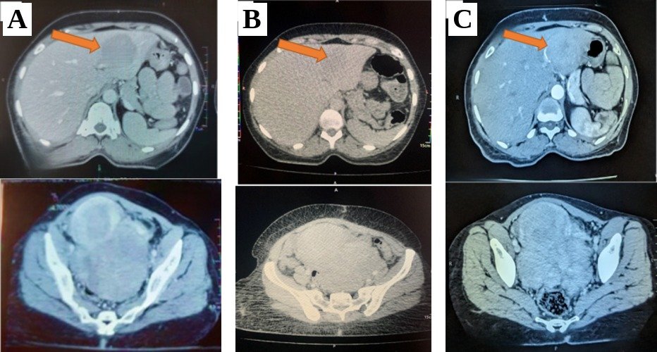
Five months after surgery, the patient was seen in the oncology clinic. She was in good general condition, with irregular hepatomegaly measuring 18 cm and a fixed pelvic mass. Thoraco-abdomino-pelvic computed tomography revealed locoregional progression with persistent peritoneal carcinomatosis and multiple secondary hepatic nodules (Figure 1A). Serum CA-125 levels were within the normal range (16.24 U/mL). The disease was staged as pT3N1M1.
After multidisciplinary discussion, first-line palliative chemotherapy with etoposide–carboplatin was initiated, based on treatment regimens extrapolated from extra-pulmonary poorly differentiated neuroendocrine carcinomas. After four cycles, tumor progression was observed, marked by an estimated 67% increase in the volume of hepatic lesions (Figure 1B), accompanied by the onset of severe abdominal pain (NRS = 8/10) requiring opioid analgesia.
Second-line chemotherapy with GEMOX (gemcitabine–oxaliplatin) was then initiated, based on reports of activity in high-grade neuroendocrine carcinomas from other primary sites. After three cycles, radiologic disease stabilization was observed (Figure 1C). Although the disease was considered stable per RECIST (8% increase in hepatic lesion volume), a third-line treatment with irinotecan–cisplatin was proposed to maintain disease control using an alternative platinum-based combination previously reported in extrapulmonary high-grade neuroendocrine tumors. This regimen was administered for six cycles, resulting in prolonged radiologic stability and partial improvement in pain symptoms. This period was complicated by repeated episodes of severe neutropenia and anemia, as well as the development of unilateral grade I hydronephrosis without significant functional impairment.
A fourth-line treatment with oral capecitabine was initiated, inspired by the CAPTEM regimen (capecitabine–temozolomide), an approach reported in neuroendocrine carcinomas, to offer a straightforward, low-intensity oral therapy in the context of cumulative hematologic toxicity, limited marrow reserve, and limited therapeutic options. It was discontinued after two cycles because of deterioration in general condition and refractory anemia requiring hospitalization. Laboratory tests at that time showed persistent anemia associated with leukocytosis and thrombocytopenia. Bone marrow examination established the diagnosis of secondary acute myeloid leukemia arising from myelodysplastic syndrome.
Given the advanced progression of the solid tumor, the deterioration in general condition, and limited bone marrow reserve, no specific treatment for leukemia was undertaken. An exclusively symptomatic and transfusion-based supportive approach was adopted.
Subsequent evolution was marked by progressive deterioration of renal function, with the development of chronic kidney disease associated with bilateral grade II hydronephrosis, as well as the occurrence of deep vein thrombosis of the lower limbs treated with anticoagulation. While awaiting nephrostomy placement, the patient’s clinical condition rapidly worsened, with the onset of encephalopathy, leading to death 23 months after the initial diagnosis.
The chronology of clinical, diagnostic, and therapeutic events is summarized in Table 1.
Table 1: Timeline of clinical, diagnostic and therapeutic events
| Time (Months) | Event |
|---|---|
| M0 | Exploratory laparotomy |
| M+1 | Pathology report: small cell neuroendocrine carcinoma of the ovary |
| M+3 | Diagnostic confirmation by immunohistochemistry (synaptophysin+, chromogranin+, cytokeratin+, Ki-67: 20%) |
| M+5 | First oncology consultation |
| M+5 to M+8 | First-line chemotherapy: etoposide–carboplatin (4 cycles) – liver tumor progression observed at the end of the protocol |
| M+9 to M+10 | Second-line chemotherapy: GEMOX (3 cycles) – 8% increase in liver volume (stable disease per RECIST) – change of treatment line initiated |
| M+11 to M+17 | Third-line chemotherapy: irinotecan–cisplatin (6 cycles) – radiological stabilization – cumulative hematological toxicities |
| M+18 | Fourth-line chemotherapy: capecitabine – rapid discontinuation due to cytopenias and deterioration of general condition |
| M+23 | Bone marrow aspirate confirming secondary acute myeloid leukemia – initiation of exclusively symptomatic/palliative care; death |
Ovarian small-cell neuroendocrine carcinomas are extremely rare, highly aggressive tumors with a strong metastatic potential, posing a major diagnostic and therapeutic challenge. Their low incidence accounts for the absence of standardized treatment recommendations and the frequent reliance on strategies extrapolated from other primary sites, such as pulmonary or gastrointestinal neuroendocrine carcinomas [5,6].
In the present case, the patient was diagnosed at an advanced stage (pT3N1M1) and received four sequential lines of chemotherapy, largely extrapolated from regimens used in advanced neuroendocrine carcinomas for other sites [7–11], with limited clinical benefit and cumulative hematologic toxicities. The first three lines were administered in the setting of aggressive, symptomatic disease, in the absence of validated therapeutic alternatives. By contrast, the use of capecitabine as fourth-line therapy was not supported by any established recommendation, reflected an approach of therapeutic exhaustion, and failed to provide any observable clinical benefit.
The occurrence of secondary acute myeloid leukemia (M+23) illustrates the high risk of hematologic toxicity following cumulative exposure to multiple cytotoxic agents in patients with ovarian cancer, particularly topoisomerase inhibitors and alkylating agents [12,13]. In this context, no specific treatment was initiated, and management remained palliative, in accordance with standards of care for patients with refractory tumors and high-risk clinical profiles. Although a temporal association between cumulative chemotherapy exposure and the development of acute leukemia was observed, no direct causal link with a specific agent can be established.
The unfavorable clinical course, tumor progression, treatment-related complications, secondary leukemia, and renal failure reflect the current limitations of available therapeutic strategies for these rare and chemoresistant tumors. The literature suggests that the overall 5-year survival for high-grade tumors at advanced stages remains very poor (9.8–21.2%), in contrast to low-grade tumors, in which it may reach up to 94% [14].
From a diagnostic standpoint, the absence of specific radiologic features for these tumors underscores the importance of systematic histopathological confirmation and the use of immunohistochemistry (synaptophysin+, chromogranin+, cytokeratin+, high Ki-67 index) for accurate diagnosis [15,16]. The relatively moderate Ki-67 index (20%) observed in this case may raise diagnostic ambiguity between a well-differentiated neuroendocrine tumor and a poorly differentiated neuroendocrine carcinoma. However, the morphological features, clinical aggressiveness, and rapid disease progression favored a diagnosis of small-cell neuroendocrine carcinoma.
This case analysis also highlights the need for a rigorous assessment of the risk–benefit ratio before initiating additional lines of chemotherapy in patients who have already been exposed to multiple regimens. Innovative approaches, such as immunotherapy or targeted therapies, may represent potential alternatives, but their evaluation is currently limited to rare case reports or small series. Zhu et al. [17] recently reported a case treated with combined immunochemotherapy using tislelizumab (anti-PD-1), etoposide, and cisplatin, achieving a rapid partial response and prolonged disease stabilization, albeit limited by severe immune-mediated adverse events. More recently, Zhao et al. [18] described a combination of carboplatin–paclitaxel, bevacizumab, and immunotherapy with tislelizumab in a patient with metastatic ovarian neuroendocrine carcinoma. This regimen resulted in a significant and durable tumor response, suggesting the potential value of an integrated strategy targeting both tumor proliferation and the immune microenvironment. Although limited, these data pave the way for prospective clinical trials and personalized treatment strategies for these rare tumors.
However, these innovative approaches must be considered within the context of resource-limited countries, particularly in sub-Saharan Africa. In Guinea, access to targeted therapies such as bevacizumab remains extremely limited due to local unavailability or prohibitive cost, and immunotherapy is not accessible in routine clinical practice. Furthermore, molecular biology analyses and immunologic characterization tests, which are essential for optimal patient selection, are not available in our setting. These structural constraints directly influenced therapeutic decisions in the present case, where management relied exclusively on conventional chemotherapy protocols. This reality highlights the persistent gap between therapeutic advances reported in the literature and their real-world applicability in resource-limited health care systems.
Similarly, the diagnosis of secondary acute myeloid leukemia was established on morphological and cytological criteria alone, in the absence of cytogenetic or molecular characterization, which is not locally available. In this context, and given the patient’s poor performance status, tumor progression, and associated comorbidities, no specific treatment for leukemia could be initiated, and an exclusively palliative approach was adopted.
This case thus illustrates the major challenges posed by rare and aggressive cancers in low-resource settings. It underscores the urgent need for adapted strategies, including more equitable access to innovative therapies, the development of international collaborations, and the strengthening of local diagnostic and therapeutic capacities, within the framework of early multidisciplinary management. In this context, early integration of palliative care and avoidance of futile chemotherapy lines appear particularly relevant to preserve quality of life and limit unnecessary iatrogenic toxicity.
This case illustrates the aggressiveness of ovarian small-cell neuroendocrine carcinomas and the limitations of conventional treatments in metastatic disease. Cumulative exposure to multiple lines of chemotherapy resulted in severe hematologic toxicities, including secondary acute leukemia, without durable oncologic benefit. It highlights the importance of a rigorous benefit–risk assessment, early integration of palliative care, and adaptation of therapeutic strategies to the realities of resource-limited settings.
Conflicts of interest: None declared.
This case report was conducted following ethical standards. Written informed consent was obtained from the patient for the publication of this case and any accompanying images. A copy of the signed consent form is available in the patient’s medical record.
*Correspondence: Ibrahima Kalil CONDÉ
condekalil1800@gmail.com
Available online: November 9, 2026
1 Department of Oncology, Donka University Hospital, Conakry, Guinea
2 Oncology and Radiotherapy Center, Hôpital du Mali, Bamako, Mali
3 Department of Hematology-Oncology, Donka University Hospital, Conakry, Guinea
© Journal of African Clinical Cases and Reviews 2026
1. Hong MK, Ding DC. Early Diagnosis of Ovarian Cancer: A Comprehensive Review of the Advances, Challenges, and Future Directions. Diagnostics (Basel). 2025;15(4):406.
2. Georgescu TA, Bohiltea RE, Munteanu O, Furtunescu F, Lisievici AC, Grigoriu C, et al. Emerging Therapeutic Concepts and Latest Diagnostic Advancements Regarding Neuroendocrine Tumors of the Gynecologic Tract. Medicina. 2021;57(12):1338.
3. Huang W, Bao Y, Luo X, Yao L, Yuan L. Neuroendocrine neoplasms of the ovary: an analysis of clinicopathological characteristics and prognosis with a focus on histological grading. Endocrine. 2022;77(1):188‑98.
4. Karpathiou G, Matias-Guiu X, Mobarki M, Vermesch C, Stachowicz ML, Chauleur C, et al. Ovarian neuroendocrine carcinoma of metastatic origin: clues for diagnosis. Human Pathology. 2019;85:309‑12.
5. He Q, Wang C, Huang D, Shen J, Liu R, Guan Y, et al. Clinicopathologic feature and treatment progress of high-grade ovarian neuroendocrine tumors. Med Oncol. 2024;42(1):2.
6. Hadoux J, Lamarca A, Grande E, Deandreis D, Kaltsas G, Janson ET, et al. Neuroendocrine neoplasms of head and neck, genitourinary and gynaecological systems, unknown primaries, parathyroid carcinomas and intrathyroid thymic neoplasms: ESMO Clinical Practice Guideline for diagnosis, treatment and follow-up. ESMO Open [Internet]. 2024 [cité 1 août 2025];9(10). Disponible sur: https://www.esmoopen.com/article/S2059-7029(24)01433-9/fulltext
7. Weaver JMJ, Hubner RA, Valle JW, McNamara MG. Selection of Chemotherapy in Advanced Poorly Differentiated Extra-Pulmonary Neuroendocrine Carcinoma. Cancers. 11 oct 2023;15(20):4951.
8. Cassier PA, Walter T, Eymard B, Ardisson P, Perol M, Paillet C, et al. Gemcitabine and oxaliplatin combination chemotherapy for metastatic well-differentiated neuroendocrine carcinomas: a single-center experience. Cancer. 2009;115(15):3392‑9.
9. Dussol A, Joly M, Vercherat C, Forestier J, Hervieu V, Scoazec J, et al. Gemcitabine and oxaliplatin or alkylating agents for neuroendocrine tumors: Comparison of efficacy and search for predictive factors guiding treatment choice. Cancer. oct 2015;121(19):3428‑34.
10. Thomas K, Voros BA, Meadows-Taylor M, Smeltzer MP, Griffin R, Boudreaux JP, et al. Outcomes of Capecitabine and Temozolomide (CAPTEM) in Advanced Neuroendocrine Neoplasms (NENs). Cancers. 2020;12(1):206.
11. Arrivi G, Verrico M, Roberto M, Barchiesi G, Faggiano A, Marchetti P, et al. Capecitabine and Temozolomide (CAPTEM) in Advanced Neuroendocrine Neoplasms (NENs): A Systematic Review and Pooled Analysis. CMAR. 2022;Volume 14:3507‑23.
12. Vay A, Kumar S, Seward S, Semaan A, Schiffer CA, Munkarah AR, et al. Therapy-related myeloid leukemia after treatment for epithelial ovarian carcinoma: An epidemiological analysis. Gynecologic Oncology. 2011;123(3):456‑60.
13. Shimada T, Saito T, Okadome M, Shimamoto K, Ariyoshi K, Eto T, et al. Secondary Leukemia After Chemotherapy and/or Radiotherapy for Gynecologic Neoplasia. International Journal of Gynecological Cancer. 2014;24(2):178‑83.
14. Pang L, Guo Z. Primary neuroendocrine tumors of the ovary: Management and outcomes. Cancer Med. 2021;10(23):8558‑69.
15. Matsutani H, Nakai G, Yamada T, Kurisu Y, Yamamoto K, Ohmichi M, et al. MRI and FDG PET/CT findings for small cell neuroendocrine carcinoma of the ovary: a case report and literature review. BMC Women’s Health. 2025;25(1):362.
16. Mantilidewi KI, Winarno GNA, Harsono AB, Suardi D, Hidayat YM, Kurniadi A, et al. Management paradigm for ovarian neuroendocrine carcinoma: a systematic review. J Ovarian Res. 2025;18:125.
17. Zhu YX, Gao XP, Xin L, Jia YC. A case report of small cell ovarian neuroendocrine carcinoma combined with immunochemotherapy. Medicine (Baltimore). 18 nov 2022;101(46):e31445.
18. Zhao Y, Kang F, Kong X, Wang N. A case report of small cell neuroendocrine carcinoma of the ovary and review of the literature. Front Immunol. 22 mai 2025;16:1569011.
